

The human brain contains roughly one hundred billion nerve cells, each of which forms thousands of connections. But how does each individual neuron know which cells to connect to and where to grow? This is one of the fundamental questions of neurobiology. Daniel Rozbeský from the Faculty of Science at Charles University and the BIOCEV centre is trying to crack it. He will now be aided in this effort by a prestigious ERC CZ grant worth 18.2 million crowns.
What is axon guidance?
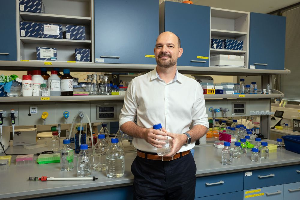
The main research focus of our laboratory. We study how axons and neurons navigate relatively complex environments during the development of the nervous system. The human brain contains approximately one hundred billion nerve cells. Each of them forms about a thousand different connections with other nerve cells. This means that we have roughly one hundred trillion connections in our heads defining our personality. They determine our memory, movement, speech, and how we respond to external stimuli – in short, they define the whole person.
A fundamental question in neurobiology is how this enormous, complicated network even comes into being. Think of it as if you had a hundred trillion cables and had to plug them into a hundred trillion sockets with absolute precision. A nerve cell has an extension called an axon. During the development of the nervous system, this axon moves and needs to get from point A to point B. But how does it do this in such a complex environment without a map?
I have no idea. How?
It needs guidance, navigation. There are signalling molecules – a kind of molecular GPS – that bind to axons. There are various receptors on the axons; the molecules bind to them, allowing information from the external environment to enter the cell. This is exactly the topic I studied during my postdoc at Oxford. I investigated what these molecules look like at the atomic level, how they bind to receptors, and how the entire signalling process is modulated.
Now we’re looking at what happens inside the cell. At certain points, the axon must decide whether to turn left, right, or go straight. Or, when an axon grows in the left hemisphere, it must decide whether to stay there or move on to the right. The fundamental question is: how does it know? The key internal structure that determines this is the cytoskeleton – the cell’s internal skeleton. Unlike the human skeleton, which is solid and rigid, the cellular cytoskeleton is extremely dynamic. It is constantly being formed and broken down.
 What exactly does this kind of cellular decision-making look like?
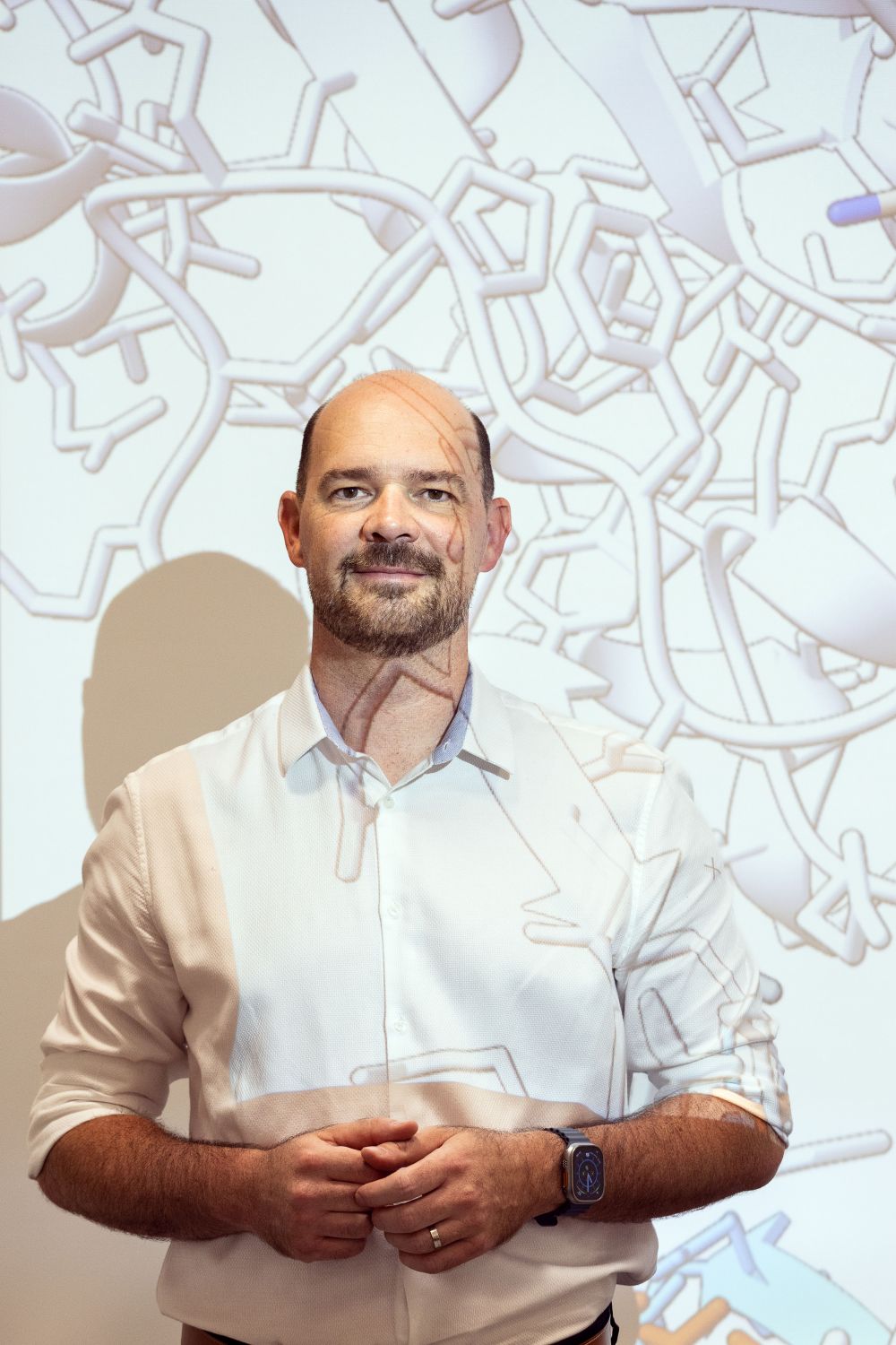
There is a structure at the end of the axon called the growth cone. It functions as a sensor. Imagine it as if you were walking in the dark and feeling around with your hands. Similarly, the axon “feels out” its surroundings with the growth cone. When it receives a signal to move in a specific direction, the formation of the cytoskeleton – specifically, actin filaments – is stimulated in that area. A large number of these filaments are produced, the cell membrane begins to stretch, and the cell grows in that direction.
Conversely, when it receives a “stop” signal – which you can imagine as bumping into a refrigerator in the dark, for example – the actin filaments begin to break down and fragment into tiny pieces. The cell membrane collapses in that direction, and the movement is halted. We’re interested in one specific protein in the lab, an enzyme called MICAL1, which we believe influences this entire process. When it’s activated, it begins to slice up the actin filaments, allowing the cell to stop growing in a specific direction.
So, this protein is what you’re mainly interested in today?
Yes – on several levels, actually. This enzyme floats freely inside the cell and must be inactivated in some way – Its activity must be turned off, otherwise it would destroy all actin filaments in its vicinity. Two years ago, we published its structure in the Nature Communications journal; this allowed us to discover how this protein is deactivated. We spent the next two years working to figure out the reverse – how it is activated. In fact, we’re currently finalising a manuscript on this very issue. We’ve managed to map out, down to the atomic level, how its activation occurs.
Another thing we’re interested in is how it binds to actin filaments and how it cuts them apart. We know from the literature that it has a unique property: it chemically modifies the filaments by oxidising them. It literally sprinkles them with oxygen atoms, causing the filament to start falling apart. We’ll be trying to understand this in detail.
You have just received an ERC CZ grant. Will you and your team be using it for further research on this particular topic?
We would like to understand how depolymerisation – i.e., actin breakdown – occurs at the molecular level. Thanks to the ERC CZ project, we will be able to move to the cellular level. We will be working directly with neurons. First, we’ll observe them using a standard fluorescence microscope and add various signalling molecules that tell them to “stop” or “go”. Since we can make the actin filaments glow, we’ll be able to see in which part of the cell they break down, what the dynamics of this process are, and how long it takes. That’s the first part of the project.
And the second part?
The first part deals with fluorescence microscopy, which provides us with information concerning dynamics. However, its drawback is resolution; we cannot get into the finest details. For that, we will use what is known as cryo-electron tomography. I am convinced that it will become the method of the 21st century in structural biology. Until now, when we wanted to determine the structure of proteins, we first had to produce and purify them or isolate them from their natural cellular environment. With X-ray crystallography, it was also necessary to convert the protein into a crystal, while in nuclear magnetic resonance it was studied dissolved in a solution, and for cryo-electron microscopy, individual protein particles are rapidly frozen in a thin layer of solution. The advantage of tomography is that we will be able to observe the structure directly inside the cells or on their surface, right within the cellular context. We will see which other structures the proteins interact with or on which parts of the membrane they are located.
It’s quite a complicated method which is still in its infancy. But I believe that in a few decades we’ll achieve very high resolution and see the actual architecture of proteins down to the atomic level. This will provide us with answers to questions that are decades, centuries, or perhaps even millennia old. It’ll be incredibly interesting to bear witness to that.
What is the principle behind cryo-electron tomography?
We rapidly freeze a living cell – in our case, a neuron. We immerse it in liquid ethane and then in liquid nitrogen, thereby immediately halting all processes inside it. We then place the cell in a cryo-electron microscope and gradually rotate it, collecting data every, say, four degrees. From these angular projections, we then calculate the 3D structure of the axon with incredible resolution – perhaps even at the nanometre level.
The disadvantage of this method, compared to fluorescence, is that by freezing we lose the time scale of the process. We stop time. Hence, we must combine both methods. We’ll link the atomic level with the movement of the entire cell while also connecting different time scales. You can imagine it as if you wanted to understand the movement of an airplane in the clouds – whether it’s flying up, down, left, or right – while at the same time looking directly into its engine to see exactly how it works.
In the third phase of the project, we will attempt to use the information we have gathered to guide axons in a targeted manner. To do this, we will use microfluidics. We will set up a maze of small chambers into which we will place signalling molecules, and we will attempt to guide the axon from point A to point B.
One of the requirements in applying for major ERC grants is to design a project that goes beyond the current boundaries of knowledge, which naturally involves significant risks. What do you think might pose challenges for your project?
I see two major risks. The first is experimental – namely, whether we will actually succeed in connecting that temporal and spatial world.
The second risk is conceptual. It may turn out that our hypothesis is wrong and that the MICAL1 protein does not play as vital a role in axon navigation as we think. Perhaps it is essential for entirely different processes, or perhaps a completely different protein – one we don’t yet know about – is actually key to navigation. But that is precisely what makes this entire project so fascinating.
Even rejecting a hypothesis brings new insight.
Exactly. Some national grants are structured so that you have to specify in your application exactly what you want to study, how you’ll go about it, what the results will be, and how it will be applied. But it’s terribly boring to map out the entire project from start to finish right at the beginning. The ERC grant is interesting precisely because you don’t know how it will turn out. We do have a strong hypothesis, but everything could turn out differently.
You’re currently working on basic research, but if we were to consider potential future applications of your project, what might they be?
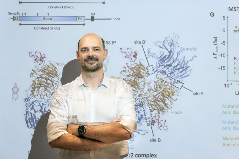
Personally, I’m mostly interested in fundamental basic research. The processes I mentioned are responsible for a wide range of pathologies, including various neurodevelopmental disorders such as autism. There are also studies pointing to a link between axon guidance and Alzheimer’s disease, and the MICAL1 protein has been associated with epilepsy and cancer. In our project, we are not primarily focused on developing treatments; rather, we want to understand the fundamental mechanisms. Only once we have a deep understanding of these processes will others be able to apply them to the development of targeted therapies.
When you were working on your ERC grant application, what helped you the most personally? And looking back, what would you have done differently?
Charles University has a very good training program for ERC grant applicants, organised primarily by Professor Zdeněk Strakoš from the Faculty of Mathematics and Physics. A few years ago, I tried applying for an ERC Starting Grant, and at that time I went through the entire preparatory cycle. I loved it. Professor Strakoš has a tremendous gift for explaining in simple terms how to write a proposal and for highlighting just how much time one needs to devote to it. Both the university’s support services and the faculty’s program for applicants are top-notch.
Feedback from colleagues was also crucial for me. I sent the project to both experts in structural biology and people completely outside my field. Based on their feedback, I kept trying to improve my application.
On the other hand, feedback can sometimes do some harm. When you send a polished project to a lot of people, there’s always someone who takes issue with something, and the advice often ends up contradicting one another. You have to know when to stop at a certain point, reflect on it, and ultimately evaluate everything yourself. Of course, I also drew on my experiences at Oxford, where I was in contact with many people who had already been successful with the ERC and provided me with their own applications as examples.
Your postdoctoral fellowship at Oxford must have been a powerful and formative experience…
It was a tough learning experience. I had a mentor and supervisor there who was extremely busy with bureaucratic tasks, which required a great deal of independence. Each team member had to design their own experiments, evaluate the results, write a paper, draft a letter to the editor, and submit the work. In the process, I encountered an overwhelming number of problems that shaped me and taught me to ask the “big questions”. And that is essential for obtaining an ERC grant – being able to ask that basic, fundamental “big question” and try to find an answer to it.
In an earlier interview, you mentioned that you enjoy “hands-on science” the most. As a team supervisor, do you still get to do any actual lab work?
These days, unfortunately, only to a very limited extent, which is a huge shame because I absolutely love it. I miss the thrill of discovery. I remember working on a project at Oxford for about three years. We needed to produce a protein, purify it, and crystallise it. Problems kept popping up, but we eventually obtained the crystals. Our goal then was to determine the structure of such a complex. The final step was collecting data from the crystals at a giant synchrotron (a particle accelerator). I remember sitting at a computer at the university around 3 a.m., waiting for the resulting dataset to arrive from the accelerator. I can still feel that tension today. When it arrived, I was so curious that I decided to quickly run all the calculations in the middle of the night. I ran the calculations on about twenty computers, and within an hour I had the first results. I could tell from them that it had worked. So I hopped on my bike and rode home, feeling satisfied. It was around six in the morning; our campus was on a little hill, and the sun was just rising, casting its light on those Oxford spires that have stood there for hundreds of years. I felt an immense sense of satisfaction. I miss that thrill and the joy of discovery a little bit today.
In 2021, you moved from Oxford to the Czech Republic, where you began setting up your own lab at the BIOCEV centre. What have the past five years been like for you?
Very challenging. First, there was the struggle to secure space, then I had to put together the entire team. Fortunately, I had decent grants – the university’s Primus and the Junior Star from the Czech Science Foundation (GA ČR). But our work is highly experimental, so I had to purchase various instruments, from chromatography systems to large centrifuges and other equipment. And that meant diving into a hellish bidding process. I understand that we’re funded by public money and that transparency is necessary, but the system constantly pushes us to buy the cheapest equipment. You can’t do cutting-edge science that way, though.
Czech science would greatly benefit from some relaxation of budgetary rules. Our grants are strictly confined to categories such as personnel expenses, materials, travel expenses… Shifting funds between these areas is a huge problem. That’s why I’d like to highlight the Primus program, which was wonderfully open, flexible, and yet completely transparent. If all national grants worked this way, it would be fantastic.
We’re also limited by the duration of the funding. These grants are usually designed to last three years, but in my field, you can’t answer a “big question” in just three years. If they were set up for five years, that would help us tremendously.
So the first few years back in the Czech Republic were challenging; it wasn't until three years later that we finally began to deliver real results, the team settled in, and only now are we beginning to reap the true rewards of our work.
You’re actually from Trnava, Slovakia. Have you ever thought about setting up a research group in your home country?
Back in Oxford, my wife and I considered whether to stay there or move to the Czech Republic or Slovakia, where we’re both from. After Brexit, however, the situation in Britain changed dramatically. A huge number of people left for continental Europe, and then COVID hit. When I talk to people from Oxford today, I hear how that relaxed and creative international atmosphere has faded a bit due to all these circumstances.
We also considered Slovakia, but the political and financial situation in science there is generally very poor. Over the past twenty years, there has been a massive exodus of intellectuals, and the funding allocated to science there is nowhere near what it is in the Czech Republic. Unfortunately, I also believe that the situation in Slovakia is unlikely to improve anytime soon.
Let us not end on such a depressing note. Which scientific achievement to date do you value the most?
I enjoy following the paths my gifted students take. For example, I had a very talented master’s student who got a “taste” of various methods and approaches to scientific thinking here. Today, he’s building on those skills at the world-class Max Delbrück Center in Berlin, where he’s doing very well. Similarly, I had a brilliant postdoc working with me who is now at a research institute in Okinawa. Uncovering how molecular mechanisms work is wonderful and I find it incredibly enjoyable, but being able to pass on my knowledge to someone and then watch their potential grow is, for me, the greatest success of all.
| Daniel Rozbeský, PhD |
| Graduated in biochemistry from the Faculty of Science at Charles University, where he also successfully defended his dissertation. He spent seven years as a postdoctoral fellow in Yvonne Jones’s group at the University of Oxford. In 2021, he returned to the Czech Republic, where he established his own laboratory focused on structural neurobiology (the Rozbesky Lab) at the BIOCEV centre. Currently, he also works at the Department of Cell Biology at the Faculty of Science, Charles University, and the Institute of Molecular Genetics of the Czech Academy of Sciences. His research focuses on the structural biology of signalling molecules and cell surface receptors, which play a key role in neurobiology and human diseases. In the past, Daniel Rozbeský received a Charles University Primus grant and a Junior Star grant from the Czech Science Foundation. This year, he applied for an ERC Consolidator Grant. Although the European Research Council’s evaluation panel commended his project, he did not receive European funding due to budget constraints. However, he subsequently received support from the Czech Ministry of Education, Youth, and Sports, and was awarded a national ERC CZ grant of 18.2 million crowns for a period of two years. |